Do Centrioles appear in meiosis or mitosis
Every animal-like cell has two small organelles called centrioles. They are there to help the cell when it comes time to divide. They are put to work in both the process of mitosis and the process of meiosis. You will usually find them near the nucleus but they cannot be seen when the cell is not dividing.
Do meiosis have centrioles?
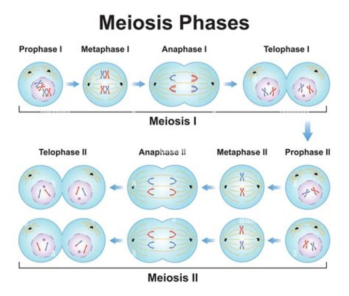
The meiosis I spindle poles contain two centrioles capable of producing two centrosomes by splitting, leading to the formation of a bipolar spindle in meiosis II division. Since the centrioles do not replicate before meiosis II division, each meiotic II spindle pole possesses only one centriole (Fig. 2G).
During which phase of mitosis do centrioles appear?
Prophase is the first stage of mitosis, during which the cell begins to position itself in order to separate the chromatids and divide. During prophase, the nuclear envelope and nucleolus are dissolved and the chromosomes condense. The centrioles and spindle fibers begin to form at opposite poles of the cell.
Are centrioles involved in mitosis?
Found only in animal cells, these paired organelles are typically located together near the nucleus in the centrosome, a granular mass that serves as an organizing center for microtubules. … Though centrioles play a role in the mitosis of animal cells, plant cells are able to reproduce without them.What do centrioles do during cell division?
The main function of centrioles is to produce cilia during interphase and the aster and the spindle during cell division.
Why do plants not need centrioles for mitosis or meiosis?
Plant cells are still able to divide without centrioles because the spindle fibers form outside the nuclear envelope. Spindle fibers are important for…
What happens to centrioles during meiosis?
When the time comes for cell division, the centrioles will appear and move to opposite ends of the nucleus. During division you will see four centrioles. One pair moves in each direction. … During prophase, the centrioles move to opposite ends of the nucleus and a mitotic spindle of threads begins to appear.
How do centrioles organize microtubules?
Centrioles are best known for their role in centrosomes, structures that act as microtubule organizing centers in animal cells. A centrosome consists of two centrioles oriented at right angles to each other, surrounded by a mass of pericentriolar material, which provides anchoring sites for microtubules.Where are the centrioles in the cell?
Centrioles are paired barrel-shaped organelles located in the cytoplasm of animal cells near the nuclear envelope. Centrioles play a role in organizing microtubules that serve as the cell’s skeletal system.
Do centrioles move to the poles?In animal cells, the centrioles near the nucleus begin to separate and move to opposite poles (sides) of the cell. As the centrioles move, a spindle starts to form between them.
Article first time published onIs cytokinesis part of mitosis?
Cytokinesis is the final physical cell division that follows telophase, and is therefore sometimes considered a sixth phase of mitosis. All phases of mitosis, as well as the flanking periods of interphase and cytokinesis before and after, are shown in Figure 8.
How do centrioles and cilia work together?
Cilia and flagella are organized from centrioles that move to the cell periphery. … Basal bodies control the direction of movement of the cilia. This can be shown experimentally. Centrioles control the direction of cilia or flagella movement.
What role do centrioles play in spindle formation in animal cells?
Centrioles are responsible for organizing the spindle fibers in the mitotic spindle apparatus and are thought to participate in the completion of cytokinesis during the process of cell division.
Do spindle fibers form in mitosis or meiosis?
During mitosis, the spindle fibers are called the mitotic spindle. Meanwhile, during meiosis, the spindle fibers are referred to as the meiotic spindle. At the beginning of nuclear division, two wheel-shaped protein structures called centrioles position themselves at opposite ends of the cell forming cell poles.
How do centrioles form cilia and flagella?
Centrioles. A basal body is a centriole, which is a cylinder-shaped structure composed of microtubules that in turn contain up to 13 protofilaments surrounding a hollow center. Basal bodies are the organelles needed to form cilia and flagella. The protofilaments are polymers of the protein tubulin.
Which of the following occurs during mitosis?
During mitosis, a eukaryotic cell undergoes a carefully coordinated nuclear division that results in the formation of two genetically identical daughter cells. … Then, at a critical point during interphase (called the S phase), the cell duplicates its chromosomes and ensures its systems are ready for cell division.
How does mitosis occur without centrioles?
plant cells without centriole build special vesicles from their Golgi apparatus which are important for cell division. In some cases the cells walls themselves organize many microtubules that form the spindle during mitosis.
How does cell division occur in plants without centrioles?
Land plants have an anastral mitotic spindle that forms in the absence of centrosomes, and a cytokinetic apparatus comprised of a predictive preprophase band (PPB) before mitosis and a phragmoplast after mitosis. … Phragmoplast development appears similar in the three taxa and to vascular plants as well.
What is the importance of the centrioles in cell division How does the plant cell compensate for its absence?
In the cell, centrioles aid in cell division by facilitating the separation of chromosomes. For this reason, they are located near the nucleus. Apart from cell division, centrioles are also involved in the formation of cilia and flagella and thus contribute to cell movement.
Where do centrioles come from?
Centrioles occur as paired cylindrical organelles together with pericentriolar material (PCM) in the centrosome of an animal cell. Centrioles are found as single structures in cilia and flagella in animal cells and some lower plant cells. Centrioles are constructed of microtubules.
Are centrioles present in prokaryotic cells?
That means, the Prokaryotic cells don’t have a centriole because prokaryotic cells have naked genetic material, not enveloped by the nuclear membrane. Centrosome is an organelle usually containing two cylindrical structures called centrioles.
Are centrioles prokaryotic or eukaryotic?
Centrioles are found in most eukaryotic cells. They are cylindrical shaped organelle largely composed of a protein called tubulin and are mainly involved in cell division and in the formation of spindle fibres.
What is the role of the Centriole in cell division quizlet?
The main function of the centriole is to help with cell division in animal cells. The centrioles help in the formation of the spindle fibers that separate the chromosomes during cell division (mitosis).
What do centrioles and basal bodies do?
Centrioles, from which basal bodies are derived, act as anchoring sites for proteins that in turn anchor microtubules, and are known as the microtubule organizing center (MTOC). These microtubules provide structure and facilitate movement of vesicles and organelles within many eukaryotic cells.
What organelles work with centrioles?
In dividing cells, they recruit a collection of proteins (known as pericentriolar material) to form larger organelles called centrosomes that nucleate microtubules and organize the spindle poles during cell division (Fu et al., 2015; Figure 1A) In non-dividing cells, centrioles are involved in the production of cilia, …
In which phase centrioles move towards opposite ends?
The centriole begins to move towards opposite poles of the cell in prophase.
Is cytokinesis part of meiosis or mitosis?
Cytokinesis is the physical process of cell division, which divides the cytoplasm of a parental cell into two daughter cells. It occurs concurrently with two types of nuclear division called mitosis and meiosis, which occur in animal cells.
What structure is produced when protein fibers radiate from Centrioles?
What structure is produced when protein fibers radiate from centrioles? Spindle fiber. What forms across the center of a cell near the end of telophase?
Does cytokinesis occur after meiosis 1?
Meiosis I ends when the chromosomes of each homologous pair arrive at opposing poles of the cell. The microtubules disintegrate, and a new nuclear membrane forms around each haploid set of chromosomes. The chromosomes uncoil, forming chromatin again, and cytokinesis occurs, forming two non-identical daughter cells.
How do centrioles move?
In animal cells, the centrioles near the nucleus begin to separate and move to opposite poles (sides) of the cell. As the centrioles move, a spindle starts to form between them. The spindle consists of fibers made of microtubules that pull chromosomes apart during cell division.
How are microtubules used in mitosis?
Microtubules play an important role in cell division by contributing to the formation of the mitotic spindle, which plays a part in the migration of duplicated chromosomes during anaphase. … The two poles of the spindle, made from microtubule structures, help to segregate and separate duplicated chromosomes reliably.