Is the C1 vertebra the axis
The C1 and C2 vertebrae are the first two vertebrae of the cervical spine. They are also called the atlas and axis vertebrae.
What is atlas and axis?
The atlas is the first cervical (neck) vertebra which is just under the head; it is named for Atlas, the Greek god who supported the world on his shoulders. The axis is the second cervical vertebra; it has what is called the odontoid process about which the atlas rotates. … It allows the head turn from side to side.
What is C1 vertebra called?
Atlas (C1) The atlas is ring-shaped and does not have a body, unlike the rest of the vertebrae. Fused remnants of the atlas body have become part of C2, where they are called the odontoid process, or dens.
What is atlas vertebra?
atlas: the first cervical vertebra (C1), lying directly under the skull, through which the head articulates with the neck. The main connection to the vertebra below is a pivot around the odontoid process that is an upward projection of the body of the second cervical vertebra.What is the name of the C2 vertebra?
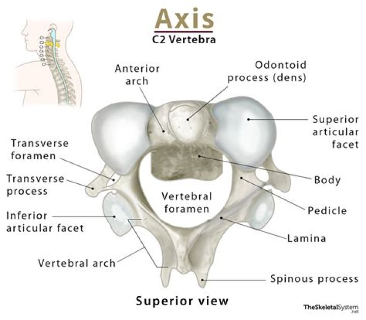
C2 Vertebra (the axis). The second vertebra, called the axis, has a large bony protrusion (the odontoid process) that points up from its vertebral body and fits into the ring-shaped atlas above it. The atlas is able to rotate around the axis, forming the atlantoaxial joint.
Is the dens anterior or posterior?
The dens or odontoid process exhibits a slight constriction or neck, where it joins the body. On its anterior surface is an oval or nearly circular facet for articulation with that on the anterior arch of the atlas.
Is there a disc between C1 and C2?
There is no intervertebral disc between C1 and C2, which is unique in the spine.
What is sacral vertebra?
The sacral vertebrae—also called the sacral spine—consists of five sacral vertebrae bones. These bones fuse together to form the sacrum, the shield-shaped bony structure located at the base of the lumbar vertebrae (the five cylindrical bones forming the spine of the lower bank) and connected to the pelvis.What does the C1 and C2 vertebrae do?
The C1 and C2 vertebrae function together to give your head flexibility. With the atlas and axis relationship, you are able to swivel and rotate your head, as well as support your head. These two vertebrae are more responsible for the head’s rotational range of motion than from any other joint.
What is spiral cord?A column of nerve tissue that runs from the base of the skull down the center of the back. It is covered by three thin layers of protective tissue called membranes. The spinal cord and membranes are surrounded by the vertebrae (back bones).
Article first time published onWhere is the C7 vertebrae?
The 7th cervical (C7) vertebra is the largest and most inferior vertebra in the neck region. Unlike the other cervical vertebrae, the C7 has a large spinous process that protrudes posteriorly toward the skin at the back of the neck.
What is the name of the first vertebra?
The first vertebra (C1) is the ring-shaped atlas that connects directly to the skull. This joint allows for the nodding or “yes” motion of the head. The second vertebra (C2) is the peg-shaped axis, which has a projection called the odontoid, that the atlas pivots around.
Why is the axis bone called the axis?
It is called the “axis” because the uppermost cervical vertebra (called the atlas) rotates about the odontoid process of C2. The joint between the axis and atlas is a pivot type of joint. … The Latin word “axis” means axle or pole. The axis bone serves as the axle about which the atlas (and the head) turn.
What is the main axis of the body?
Explanation: In anatomy, the second cervical vertebra (C2) of the spine is named the axis (from Latin axis, “axle”) or epistropheus. By the atlanto-axial joint, it forms the pivot upon which the first cervical vertebra (the atlas), which carries the head, rotates.
What are the 4 types of vertebrae?
There are 33 vertebrae in the human spine that are split into four regions that correspond to the curvature of the spine; the cervical, thoracic, lumbar, sacrum, and coccyx.
Where is the C4 and C5 vertebrae?
The C3, C4, and C5 vertebrae form the midsection of the cervical spine, near the base of the neck. A cervical vertebrae injury is the most severe of all spinal cord injuries because the higher up in the spine an injury occurs, the more damage that is caused to the central nervous system.
How many axis vertebrae are there?
You’re born with 33, but the sacrum and coccyx fuse to the rest of the spine, making it 24 by the time you’re an adult. Of those 24 (not counting the sacrum and coccyx), two vertebrae are fortunate enough to have names. The atlas (C01) and axis (C02) are two of the most important vertebrae in the spine.
Where is the C5 and C6 vertebrae located?
The C5-C6 spinal motion segment (located in the lower cervical spine just above the C7 vertebra) provides flexibility and support to much of the neck and the head above.
What are the 7 vertebrae?
It consists of 7 bones, from top to bottom, C1, C2, C3, C4, C5, C6, and C7. In tetrapods, cervical vertebrae (singular: vertebra) are the vertebrae of the neck, immediately below the skull. Truncal vertebrae (divided into thoracic and lumbar vertebrae in mammals) lie caudal (toward the tail) of cervical vertebrae.
How do you know if your neck is out of alignment?
- chronic pain.
- joint stiffness.
- slouched posture.
- reduced range of motion.
- decreased mobility.
- discomfort when sitting, standing, and laying down.
- permanent joint and bone deformities.
- broken bones, especially in the spine.
What happens if you break your C2 vertebrae?
C1 and C2 Vertebrae Breaks, Fractures, and Misalignments Symptoms following an injury to the cervical vertebrae C1 and C2 may include: Complete paralysis of arms and legs. Muscle atrophy. Limited head and neck movement.
What happens when C1 is out of alignment?
The natural postural reflex gets overridden. Now what happens is because C0-C1-C2 is the most freely movable joint in the spine, then we get a rotational malposition, or misalignment of C1. This malposition or misalignment starts to cause problems in how the head and the neck talk to each other.
Does the axis have transverse foramen?
The axis. The second cervical vertebra is the axis (Fig. … Both transverse processes have a transverse foramen for the vertebral arteries. The superior articular facets of the axis articulate with the inferior articular facets of the atlas.
Does the axis have a vertebral arch?
The axis is ossified from five primary and two secondary centres. The body and vertebral arch are ossified in the same manner as the corresponding parts in the other vertebrae, viz., one centre for the body, and two for the vertebral arch.
Which vertebrae has no vertebral?
The atlas (C1 vertebra) does not have a body or spinous process. It consists of an anterior and a posterior arch and elongated transverse processes.
What does C1 and C2 control?
C1, C2, and C3 (the first three cervical nerves) help control the head and neck, including movements forward, backward, and to the sides. 1. The C2 dermatome handles sensation for the upper part of the head, and the C3 dermatome covers the side of the face and back of the head. 2. (C1 does not have a dermatome.)
Can you survive a C1 break?
Injuries to the C1 and C2 vertebrae are rare, accounting for only 2% of spinal injuries each year. However, they are also considered to be the worst spinal cord injury that it is possible to sustain, and often fatal.
What is the coccyx?
What is the tailbone/coccyx? Your coccyx is made up of three to five fused vertebrae (bones). It lies beneath the sacrum, a bone structure at the base of your spine. Several tendons, muscles and ligaments connect to it.
What vertebral level is the coccyx?
The coccyx, commonly known as the tailbone, is below the sacrum. Individually, the sacrum and coccyx are composed of smaller bones that fuse (grow into a solid bone mass) together by age 30. The sacrum is made up of 5 fused vertebrae (S1-S5) and 3 to 5 small bones fuse creating the coccyx.
Where is S3 in the spine?
The sacral spinal nerve 3 (S3) is a spinal nerve of the sacral segment. It originates from the spinal column from below the 3rd body of the sacrum.
Why does L2 end spinal cord?
It is these spinal nerve roots that compose the cauda equina beyond L1/L2. The fact that the spinal cord ends at L1/L2 is very useful in clinical practice in that it allows for spinal taps to be performed to sample CSF without the risk of puncturing the spinal cord.