What is an electrode in ECG
Electrodes (small, plastic patches that stick to the skin) are placed at certain spots on the chest, arms, and legs. The electrodes are connected to an ECG machine by lead wires. The electrical activity of the heart is then measured, interpreted, and printed out. No electricity is sent into the body.
What are the electrodes used in ECG?
Two types of electrodes in common use are a flat paper-thin sticker and a self-adhesive circular pad. The former are typically used in a single ECG recording while the latter are for continuous recordings as they stick longer.
Why are electrodes placed in ECG?
It is important an ECG is recorded accurately. ECG electrode placement is standardised, allowing for the recording of an accurate trace – but also ensuring comparability between records taken at different times.
What are leads and electrode?
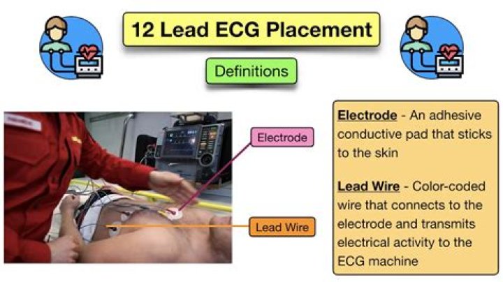
An electrode is a conductive pad that is attached to the skin and enables recording of electrical currents. An ECG lead is a graphical description of the electrical activity of the heart and it is created by analysing several electrodes.Where are electrodes placed for ECG?
To properly record a 12-lead ECG, it is important to have the patient lying comfortably with the wrist close to but not touching the trunk. The limb electrodes should be placed on the right and left wrists and the right and left ankle.
What are the electrodes used for ECG EEG and EMG measurement?
Surface Ag/AgCl electrodes are the most common and favoured electrodes in clinical measurements for recording biological signals such as ECG, EMG and EEG [16]. One of the main advantages of using Ag/AgCl electrodes is the low noise level it generates during biological signals recording [16].
Which electrode is used for EEG?
Wet/Gel electrodes Also, low electrode-skin impedance can help to reduce the power line interference and also make EEG signals more immune to movement artifacts, including cable motion. Today, the most commonly used electrode is the silver/silver chloride (Ag/AgCl) electrode.
How many electrodes are on an ECG?
Although it is called a 12-lead ECG, it uses only 10 electrodes. Certain electrodes are part of two pairs and thus provide two leads.What is the purpose of the electrode?
Electrodes are used to provide current through nonmetal objects to alter them in numerous ways and to measure conductivity for numerous purposes. Examples include: Electrodes for fuel cells.
What is aVL lead in ECG?aVL means augmented Vector Left; the positive electrode is on the left shoulder. aVF means augmented Vector Foot; the positive electrode is on the foot.
Article first time published onHow many electrodes will you place on your subject for today's activity if you use a standard ECG apparatus?
During a resting ECG, a technician will attach 10 electrodes with adhesive pads to the skin of your chest, arms and legs. Men may have chest hair shaved to allow a better connection. You will lie flat while the computer creates a picture, on graph paper, of the electrical impulses traveling through your heart.
How do you place a 3 lead ECG electrode?
- WHITE.
- RA (right arm), just below the right clavicle.
- BLACK.
- LA (left arm), just below the left clavicle.
- RED.
- LL (left leg), on the lower chest, just above and left of the umbilicus.
Why should the expiration date on an electrode pouch be checked?
Each electrode pouch (and the box containing the pouches) is stamped with an expiration date. The medical assistant must always check the expiration date of the electrodes before applying them. The electrolyte gel on outdated electrodes may be dried out; a dried out electrolyte is unable to transmit a good ECG signal.
What is electrode placement?
Electrode Placement. Electrode Placement and Lead Selection. Proper electrode placement is essential to acquire accurate ECG strips. ECG monitor manufacturers provide safe use instructions that will include electrode placement guidelines specific to their products.
How do you place electrodes on a 12 lead ECG?
- Prepare the skin. …
- Find and mark the placements for the electrodes:
- First, identify V1 and V2. …
- Next, find and mark V3 – V6. …
- Apply electrodes to the chest at V1 – V6. …
- Connect wires from V1 to V6 to the recording device. …
- Apply limb leads.
Why are electrodes applied to the fleshy part of a limb?
why place electrodes on fleshy parts of the limbs? to reduce artifacts. the myocardial cells recover electrically.
How do EEG electrodes work?
During an EEG, small electrodes and wires are attached to your head. The electrodes detect your brain waves and the EEG machine amplifies the signals and records them in a wave pattern on graph paper or a computer screen (Fig. … A sample EEG recording showing a focal spike typical of a seizure.
How do you identify electrodes?
The letter “E” indicates an electrode. The first two digits represent the resulting weld’s minimum tensile strength, measured in pounds per square inch (psi). For example, the number 70 in a E7018 electrode indicates that the electrode will produce a weld bead with a minimum tensile strength of 70,000 psi.
How are EEG electrodes attached?
A technician attaches discs (electrodes) to your scalp using a special adhesive. Sometimes, an elastic cap fitted with electrodes is used instead. The electrodes are connected with wires to an instrument that amplifies the brain waves and records them on computer equipment.
What do EMG electrodes detect?
Electromyography (EMG) measures muscle response or electrical activity in response to a nerve’s stimulation of the muscle. The test is used to help detect neuromuscular abnormalities. During the test, one or more small needles (also called electrodes) are inserted through the skin into the muscle.
What is needle electrodes?
Definition: needle electrode. needle electrode. A fine wire through which electrical current may flow when attached to a power source; used to carry high frequency electrical currents that create heat or destroy diseased tissue (called radiofrequency ablation) or seal blood vessels.
What is electrode surface?
A small device that is attached to the skin to measure or cause electrical activity in the tissue under it. Surface electrodes may be used to look for problems with muscles and nerves.
WHAT IS A electrode in medical terms?
(ee-LEK-trode) In medicine, a device such as a small metal plate or needle that carries electricity from an instrument to a patient for treatment or surgery. Electrodes can also carry electrical signals from muscles, brain, heart, skin, or other body parts to recording devices to help diagnose certain conditions.
How does a medical electrode work?
Medical electrodes proceed with quantification of internal ionic currents and results in diagnosis of various ocular, nervous, cardiac, and muscular disorders. … The device works through provision of an electrical contact between apparatus used to monitor activities and patient.
What are the 3 types of electrode?
The three primary categories of electrodes—cellulose, rutile, and basic (low-hydrogen)—obtain their name from the coating type.
How many electrodes are used in EEG?
Typically EEG systems with 64 or more electrodes are used. However, in practical applications, set-ups with fewer electrodes are required.
How often should you replace the electrodes?
Electrodes should be changed daily. Electrode placement is integral for accurate results. When an electrode is misplaced by as little as one intercostal space, QRS morphology may change and contribute to misdiagnosis.
How many limb electrodes are in a standard ECG?
The standard ECG has 12 leads. Six of the leads are considered “limb leads” because they are placed on the arms and/or legs of the individual. The other six leads are considered “precordial leads” because they are placed on the torso (precordium). The six limb leads are called lead I, II, III, aVL, aVR and aVF.
What does V1 V2 V3 mean in ECG?
The areas represented on the ECG are summarized below: V1, V2 = RV. V3, V4 = septum. V5, V6 = L side of the heart. Lead I = L side of the heart.
Are leads 1 and aVL contiguous?
These leads view the high lateral wall of the left ventricle. I & aVL Lateral Wall Leads V5 and V6 are positioned on the left lateral chest and view the lower lateral wall of the left ventricle. Since Leads 1, aVL, V5 and V6 all view the lateral wall of the left ventricle they are considered contiguous.
What are the 3 bipolar leads?
The bipolar extremity leads are called I, II and III. The unipolar extremity leads are called avR, avL and avF, and the chest leads are called V1–V6.