What is CTDI volume in CT
The CTDIvol specifies the radiation intensity used to perform a specific CT examination and is the metric used by the American College of Radiology (ACR) for CT practice accreditation (20). For identical radiographic techniques (same kilovolt peak and milliampere second), CTDIvol values differ by about a factor of two.
What is the purpose of CTDI?
The CTDI is a simple, standardized measure of the dose output of a CT scanner that can be used to compare different scan techniques on a single scanner or between scanners. CTDI is an averaged dose to a standard phantom from a mul- tiple scan examination where the patient table is incremented between scans.
What is the unit of the CTDI?
The computed tomography dose index (CTDI) is a commonly used radiation exposure index in X-ray computed tomography (CT), first defined in 1981. The unit of CTDI is the gray (Gy) and it can be used in conjunction with patient size to estimate the absorbed dose.
How do you calculate CTDI volume?
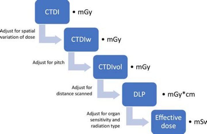
The CTDI calculation assumes that the radiation decreases linearly from the outside to the center and is calculated as CTDI = (1/3) * radiationcenter + (2/3) * radiationperiphery. It is then divided by the length of the scan to give a CTDI per slice.What is CTDI and DLP?
The term CTDI(vol), in the unit of milligray (mGy), is a reference value used for the measurement of radiation dose in a plastic cylinder. DLP stands for dose-length product. DLP is the CTDI(vol) multiplied by the scan length in centimeters and is given in units of mGy–cm.
How do you calculate DLP?
DLP is the dose length product. It is the CTDIvol multiplied by the length of the scan. The units are mGy centimeters (mGy cm). The DLP can be used to calculate a rough estimate of the effective dose.
How long does CT last?
The test will take about 30 to 60 minutes. Most of this time is spent getting ready for the scan. The actual test only takes a few minutes.
Which of the following CT phantoms is used to assess the CTDI?
These regulations specify the composition, diameter, and length of two polymethylmethacrylate (ie, acrylic, Lucite) cylindrical phantoms that are to be used for CTDI measurements. To quantify the scanner output for head CT examinations, a 16-cm-diameter phantom is to be used.What is CTDIvol in CT scan?
All modern computed tomography (CT) scanners display a value called CT dose index-volume (CTDI-vol), in units of milliGray (mGy). CTDI-vol is calculated by the scanner based on the radiation output for the particular scan.
What do we measure in CT?A computerized tomography (CT) scan combines a series of X-ray images taken from different angles around your body and uses computer processing to create cross-sectional images (slices) of the bones, blood vessels and soft tissues inside your body. CT scan images provide more-detailed information than plain X-rays do.
Article first time published onWhat is SSDE in CT?
Size specific dose estimate (SSDE) measured in mGy, is a method of estimating CT radiation dose that takes a patient’s size into account. CTDIvol and DLP are common methods to estimate a patient’s radiation exposure from a CT procedure.
What is pitch in CT?
The pitch in multislice spiral CT is defined as the ratio of the table increment over the detector collimation in this study.
What is a CTDI phantom?
CT Dosimetry Phantom. The 3-part CTDI phantom can be used with any CT system and may be used to image and monitor adult head and body, as well as paediatric dose requirements. A 2-part CTDI dosimetry phantom (adult head and body) is also available.
How many mGy is a CT scan?
A CT examination with an effective dose of 10 millisieverts (abbreviated mSv; 1 mSv = 1 mGy in the case of x-rays.) may be associated with an increase in the possibility of fatal cancer of approximately 1 chance in 2000.
Is mGy the same as mSv?
The unit milligray (mGy) is used for other types of radiation doses, but for this discussion the only one we need to know is absorbed dose. For x rays, gamma rays, and beta radiation, the conversion factor between absorbed dose in mGy and equivalent dose in mSv is one (1). So, in this case, we can say mGy equals mSv.
How do you convert mGy cm to mSv?
The conversion factors used to Convert Your Dose from Dose Length Product (mGy · cm) to Effective Dose (mSv) were 0.0022 mSv/mGy · cm for Head CT, 0.0054 mSv/mGy · cm for Neck CT and 0.0180 mSv/mGy · cm for Body CT.
What's better CT or MRI?
Both MRIs and CT scans can view internal body structures. However, a CT scan is faster and can provide pictures of tissues, organs, and skeletal structure. An MRI is highly adept at capturing images that help doctors determine if there are abnormal tissues within the body. MRIs are more detailed in their images.
Why do you need to drink water before a CT scan?
Preparing for a CT scan The water hydrates you prior to having contrast media for the CT. In the waiting area you will be asked to drink another 500ml of water which outlines the stomach and bowel clearly on the scans. The water also helps fill your bladder so that it shows on the scan.
Can you get rid of radiation from a CT scan?
Kieran Murphy, a radiologist at the university, said that a cocktail of antioxidants he and his team have developed could cut the damage done to DNA by radiation from CT scans by as much as 50%, if taken before the scan.
How many mSv are in a SV?
One sievert is 1,000 millisieverts (mSv).
How many mSv is one gray?
SI unitsHistorical dosimetry1 Gray100 R1 Sievert100 rad => 100 rem10 mGy1 Roentgen10 mSv1 rad => 1 rem
How do you calculate mSv?
Since the radiation weighting factor (WR) for γ-rays is 1, the whole body being evenly exposed to 1 mGy means that the whole body is evenly exposed to 1 mSv (1 gray × 1 (WR) = 1 millisievert). That is, equivalent doses are 1 mSv for all organs and tissues.
What is total exam DLP?
Dose Length Product (DLP) • DLP is a proxy for the total absorbed dose in a phantom over the length of a scan. • DLP is useful for comparing exam doses if scan lengths are equivalent. • DLP is measured in milligray-centimeter (mGy-cm).
How does iterative reconstruction work?
Iterative reconstruction refers to an image reconstruction algorithm used in CT that begins with an image assumption, and compares it to real time measured values while making constant adjustments until the two are in agreement.
Is a CT scan better with or without contrast?
CT of the brain can be done with or without contrast, but it is often not needed. In general, it is preferred that the choice of contrast or no contrast be left up to the discretion of the imaging physician.
Why would a doctor order a full body scan?
Why doctors order full body CT scans Detect internal injuries and bleeding. Find blood clots, tumors, and infections. Show bone fractures and muscle inflammation. Monitor diseases of the heart, liver, and lungs.
Why would a doctor order a CT scan of the chest?
CT scans of your chest can help your doctor diagnose, or rule out, various lung impairments. Some of these include blood clots, lung tumors or masses, excess fluid around the lungs (pleural effusion), emphysema, COPD, pneumonia, scarring of the lungs, tuberculosis or a pulmonary embolism.
What is helical pitch CT?
Helical CT scanning is described by defining the pitch ratio, which is the ratio of the distance moved by the table (patient) in one rotation of the x-ray tube divided by the nominal x-ray beam width. A helical scan performed using a pitch ratio of 1 corresponds most closely to contiguous axial scanning.
What does pitch affect in CT?
In the spiral CT technique, the term pitch is known, which is the distance (speed) of the table for one x-ray rotation in gantry (l) compared to the number and width of the detector (nT). Pitch changes will affect the spatial resolution of the patient’s image and dose received.
What is kVp in CT?
Kilovoltage peak (kVp) is the peak potential applied to the x-ray tube, which accelerates electrons from the cathode to the anode in radiography or computed tomography. Tube voltage, in turn, determines the quantity and quality of the photons generated.
What are CT phantoms made of?
Purpose: Currently, single-photon emission computed tomography (SPECT)/computed tomography (CT) lung phantoms are commonly constructed using polystyrene beads and interstitial radioactive water.