What is ROP
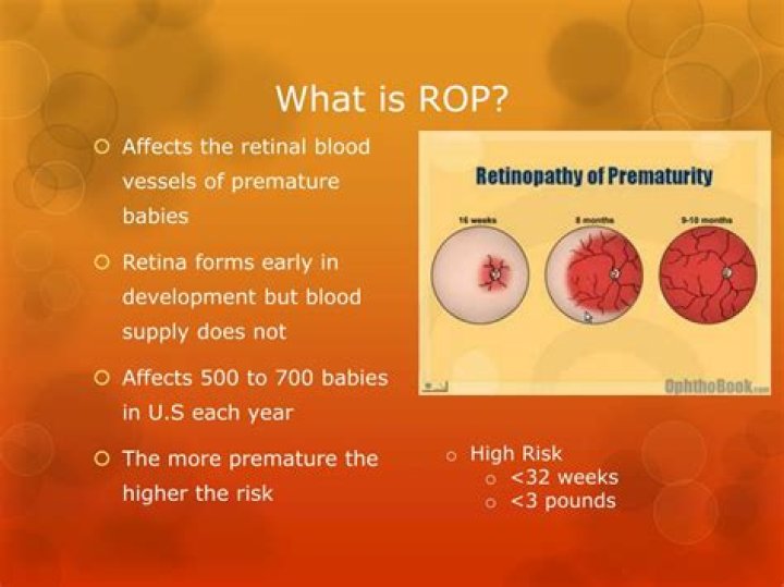
Retinopathy of prematurity (ROP) is an eye disease that can happen in premature babies. It causes abnormal blood vessels to grow in the retina, and can lead to blindness.
Is ROP curable?
In most cases, ROP resolves without treatment, causing no damage. Advanced ROP , however, can cause permanent vision problems or blindness.
What is ROP exam?
Definition. Eye exams can show the tissue and blood vessels at the back of the eye. This area is called the retina of the eye. Babies with retinopathy of prematurity (ROP) have problems with blood vessel growth in this area.
How long does ROP last?
If your child has mild retinopathy of prematurity (Stage 1 or 2), the abnormal retinal blood vessels usually heal on their own sometime in the first four months of life. But if the ROP worsens, he may need treatment.Why does ROP happen?
When children are born early, the blood vessels that feed the retina usually haven’t finished growing. ROP occurs when these vessels actually stop growing for a time, then begin growing abnormally and randomly. The new vessels are fragile and can leak, leaving the retina scarred.
What are the stages of retinopathy of prematurity?
- Stage I — Mildly abnormal blood vessel growth. …
- Stage II — Moderately abnormal blood vessel growth. …
- Stage III — Severely abnormal blood vessel growth. …
- Stage IV — Partially detached retina. …
- Stage V — Completely detached retina and the end stage of the disease.
Is ROP test painful?
Although the examination of retinopathy in premature infants is essential for identifying and improving visual acuity in a small percentage but significant number of infants, the available evidence indicates that the screening examination of ROP is usually a painful, uncomfortable, and dangerous method in the NICU (5, …
Why do preemies eyes roll back?
Doctors think it happens to preemies because the baby’s brain hasn’t developed enough to control their eye muscles. Other factors play into it as well: Brain or nerve problems like water on the brain, bleeding in the brain, seizure disorders, cerebral palsy, and other conditions. Retinal damage from ROP.What do you call a person who checks eye problem?
An ophthalmologist diagnoses and treats all eye diseases, performs eye surgery and prescribes and fits eyeglasses and contact lenses to correct vision problems. Many ophthalmologists are also involved in scientific research on the causes and cures for eye diseases and vision disorders.
When is ROP screening done?Initial screening should be performed at 31 weeks’ postmenstrual age in infants with gestational ages of 26 6/7 weeks or less at birth, and at four weeks’ chronological age in infants with gestational ages of 27 weeks or more at birth by an ophthalmologist skilled in the detection of ROP.
Article first time published onWho needs ROP screening?
Infants with a birth weight of 1,500 and 2,000 grams or a gestational age of more than 30 weeks should be screened if other health troubles put them at high risk for ROP. The first exam should occur four to nine weeks after birth, depending on how premature the baby is.
What is mild ROP?
Stage 1 is the mildest form of ROP. Babies at this stage or stage 2 often don’t need any treatment and will have normal vision. Babies with stage 3 have more blood vessels that are abnormal. These may be large or twisted, which means the retina could start to come loose.
How can ROP be prevented?
Preventing ROP before delivery A course of steroids, given to mothers likely to give birth prematurely, improves survival and reduces the complications of prematurity, including ROP. Antenatal steroids should be routine for mothers likely to give birth to a baby of less than 35 weeks’ gestation.
How is ROP screening done?
This test is done inside the neonatal unit or hospital, during a visit by the retina specialist or paediatric ophthalmologist. The pupils of the eye are dilated using drops and the retina is examined with an indirect ophthalmoscope for signs of ROP.
Can opticians prescribe?
Optometrists. … Optometrists can prescribe and fit glasses, contact lenses and low vision aids, and, if trained to do so, medicines to treat eye conditions.
Which eye care professional makes prescription glasses?
Opticians are optical professionals who fill prescriptions, issued by ophthalmologists and optometrists, for corrective eyewear. These prescriptions may include eyeglasses, contact lenses, low vision aids, and ocular prostheses.
Can optometrists diagnose eye diseases?
Optometrists can also uncover other health problems just by examining your eyes. Not only can they diagnose eye diseases but they can diagnose other diseases in the body like diabetes and hypertension.
When are preemies not considered preemies anymore?
If born between weeks 38 or 39 to 42, the baby is considered full-term. So, what is considered a premature baby? Definitions differ slightly among medical experts and organizations, but in general, when a baby is born at 37 or 38 weeks or earlier, he is considered premature, and the birth is called preterm.
Why do NICU babies have big heads?
He will be very small and his head may appear too big for his body. The reason for this is that preterm babies lack the subcutaneous fat that fills babies out in the last few weeks before birth.
Is retinopathy a disease?
Retinopathy means disease of the retina. There are several types of retinopathy but all involve disease of the small retinal blood vessels. Signs of retinopathy (see photograph) can be seen when the retina is viewed through the pupil with an ophthalmoscope.
What is NEC in preemies?
Necrotizing enterocolitis (nek-roh-TIE-zing en-ter-oh-coh-LIE-tis), or NEC, is the most common and serious intestinal disease among premature babies. It happens when tissue in the small or large intestine is injured or inflamed.
What is Stage 0 retinopathy of prematurity?
staging of retinopathy of prematurity 1,3. stage 0 – immature retinal vascularization with no demarcation line. stage 1 – thin demarcation line between vascular and avascular portions of retina. stage 2 – ridge-like structure between vascular and avascular portions of retina.