What is the falx cerebelli
The falx cerebelli is located below the tentorium cerebelli on the middle of the occipital bone. This small dural infolding extends into the space between the cerebellar hemispheres, attaching to the occipital crest of the skull and the posterior portion of the tentorium.
What is falx cerebri falx cerebelli tentorium cerebelli?
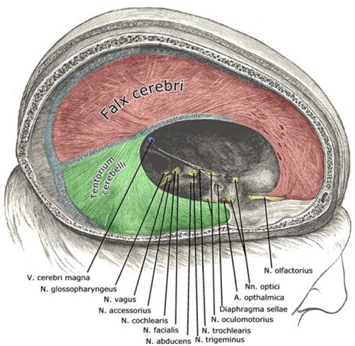
One of these, the falx cerebri, is a sickle-shaped partition lying between the two hemispheres of the brain. Another, the tentorium cerebelli, provides a strong, membranous roof over the cerebellum. A third, the falx cerebelli, projects downward from the tentorium cerebelli between the two cerebellar hemispheres.
What is the falx cerebri formed by?
Falx cerebri is a sickle-shaped vertical fold of dura that begins anteriorly at the crista galli and winds around the corpus callosum between the two cerebral hemispheres to reach the falcotentorial junction on the superior aspect of the tentorium cerebelli (Fig. 2.1A).
What is the falx cerebri and its function?
The falx cerebri separates the cerebral hemispheres and houses the dural sinuses, into which the blood and cerebrospinal fluid drain.What is the function of tentorium cerebelli?
The tentorium cerebelli functions as a partition, dispelling the burden of weight from supratentorial structures upon inferior brain matter. Clinicians and neurosurgeons, when assessing pathological findings, should have knowledge regarding the tentorium cerebelli anatomy.
What is the tentorium cerebelli made of?
The tentorium cerebelli (Latin for “tent of the cerebellum”) is an invagination of the meningeal layer of the dura mater that separates the occipital and temporal lobes of the cerebral hemispheres from the cerebellum and brainstem.
What are dural folds?
The meningeal layer of the dura mater creates several dural folds that divide the cranial cavity into freely communicating spaces. The function of the dural folds is to limit the rotational displacement of the brain. The folds include the following: The falx cerebri is a meningeal projection of dura in the brain.
What is the arachnoid mater made of?
The arachnoid is composed of collagen and elastic fibers. It has a variable thickness, in places being formed by several cell layers. Its outer (dural) aspect is smoother than the inner (pial) aspect from which trabeculae emerge to bridge the subarachnoid space (Nicholas and Weller, 1988).What is the FALX Cerebelli made up of?
Anatomical terms of neuroanatomy The falx cerebri, also known as the cerebral falx, is a large, crescent-shaped fold of meningeal layer of dura mater that descends vertically in the longitudinal fissure between the cerebral hemispheres of the human brain.
What is the meaning of falx?a Latin word meaning “sickle” (= a tool with a curved C-shaped blade), used in the names of body parts that are shaped like a sickle: the cerebral falx (= part of the brain shaped like a C or sickle)
Article first time published onWhat separates the two hemispheres of the cerebrum?
A fissure or groove that separates the two hemispheres is called the great longitudinal fissure. The two sides of the brain are joined at the bottom by the corpus callosum. The corpus callosum connects the two halves of the brain and delivers messages from one half of the brain to the other.
What is falx tentorium?
Here’s the tentorium. Its full name is tentorium cerebelli. It separates the posterior cranial fossa from the rest of the cranial cavity, and separates two major parts of the brain, the cerebrum above from the cerebellum below. … The falx forms a mid-line partition between the two cerebral hemispheres.
What is in the circle of Willis?
The Circle of Willis is the joining area of several arteries at the bottom (inferior) side of the brain. At the Circle of Willis, the internal carotid arteries branch into smaller arteries that supply oxygenated blood to over 80% of the cerebrum.
What is the singular form of meninges?
meninges, singular meninx, three membranous envelopes—pia mater, arachnoid, and dura mater—that surround the brain and spinal cord.
What does FALX Cerebelli separate?
The falx cerebri divides the two cerebral hemispheres, while the tentorium separates the cerebral lobes from the underlying cerebellum (figure 1).
What is inferior to the tentorium cerebelli?
The tentorium cerebelli is attached to the falx cerebri at its midline, and this attachment contains the straight sinus. … This margin lies inferior to the tentorial notch and extends anteriorly to attach to the posterior clinoid process and contains the superior petrosal sinus 1.
What is true about crus cerebri?
The cerebral crus (crus cerebri) is the anterior portion of the cerebral peduncle which contains the motor tracts, travelling from the cerebral cortex to the pons and spine. The plural of which is cerebral crura.
What is dura in spine?
The dura is a thin layer of tissue that covers and protects the spinal cord. It lies in between the spine (the bone) and the spinal cord (nerve tissue). Durotomy, which is an incision in the dura, can be a planned part of the surgical technique.
Why is it called dura mater?
The name dura mater derives from the Latin for tough mother (or hard mother), a loan translation of Arabic أم الدماغ الصفيقة (umm al-dimāgh al-ṣafīqah), literally ‘thick mother of the brain’, matrix of the brain, and is also referred to by the term “pachymeninx” (plural “pachymeninges”).
What is dura of the brain?
Listen to pronunciation. (DER-uh MAY-ter) The tough outer layer of tissue that covers and protects the brain and spinal cord and is closest to the skull. The dura mater is one of the three layers that form the meninges.
Where is the supratentorial compartment?
The supratentorial area (the upper part of the brain) contains the cerebrum, lateral ventricle and third ventricle (with cerebrospinal fluid shown in blue), choroid plexus, pineal gland, hypothalamus, pituitary gland, and optic nerve.
What is superior to tentorium cerebelli?
The falx cerebri (a vertical fold of dura and the largest of the four dural septa) attaches to the superior surface of the tentorium cerebelli along the midline (the straight sinus is contained within this attachment), and a further vertical fold of dura; the falx cerebelli is attached to its inferior surface along its …
Where does the tentorium attach?
The tentorium is a tent-shaped duplicated fold of meningeal dura that attaches to the sphenoid bone anteriorly, the petrous temporal bone laterally, and the squamous part of the occipital bone posteriorly.
What is posterior fossa?
The posterior fossa is a small space in the skull, found near the brainstem and cerebellum. The cerebellum is the part of the brain responsible for balance and coordinated movements. The brainstem is responsible for controlling vital body functions, such as breathing.
What is the superior sagittal sinus?
The superior sagittal sinus is an unpaired venous structure that originates at the junction of the frontal and ethmoid bone, directly posterior to the foramen cecum close to the crista galli.
What is the GREY matter?
Anatomical terminology. Grey matter (or gray matter) is a major component of the central nervous system, consisting of neuronal cell bodies, neuropil (dendrites and unmyelinated axons), glial cells (astrocytes and oligodendrocytes), synapses, and capillaries.
What is spiral cord?
A column of nerve tissue that runs from the base of the skull down the center of the back. It is covered by three thin layers of protective tissue called membranes. The spinal cord and membranes are surrounded by the vertebrae (back bones).
What type of tissue is pia mater?
The term “pia mater” means “tender matter.” It is composed of delicate connective tissue and has many tiny blood vessels. The pia mater is the only layer that clings tightly to the brain and follows all of its convolutions. Cerebral arteries and veins travel in the subarachnoid space, completely enveloped by pia mater.
What does cerebri mean in medical terms?
Cerebral: Of or pertaining to the cerebrum or the brain.
What is midline falx pregnancy?
The midline falx is visible as an echogenic line running anteroposteriorly in the midline bisecting the “butterfly” (Fig. 1.6). The posterior fossa contains the developing cerebellum.
What is the difference between gyri and sulci?
Gyri and sulci are the folds and indentations in the brain that give it its wrinkled appearance. Gyri (singular: gyrus) are the folds or bumps in the brain and sulci (singular: sulcus) are the indentations or grooves.